Thank you very much. I think I’ll work more on this topic.
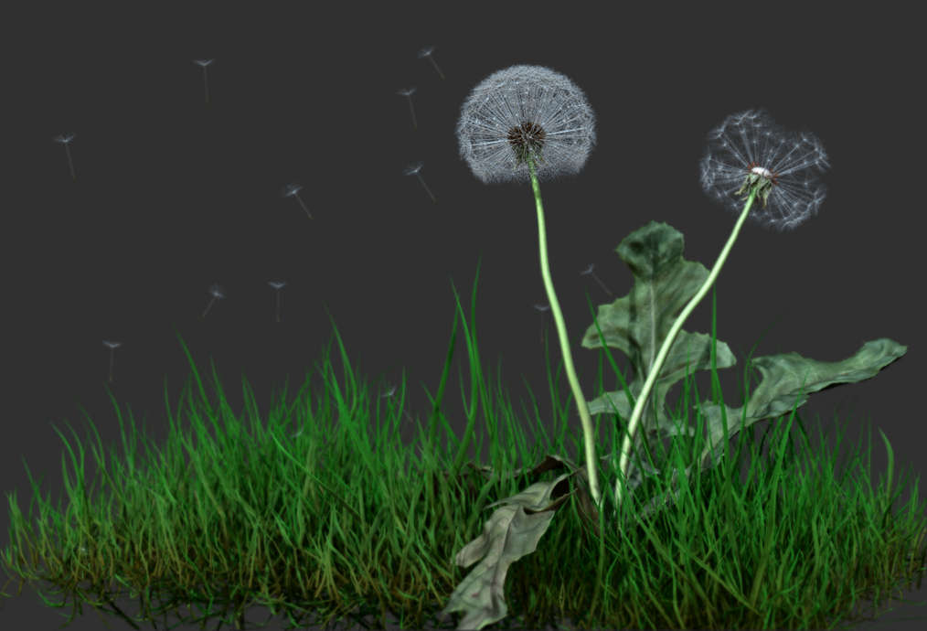
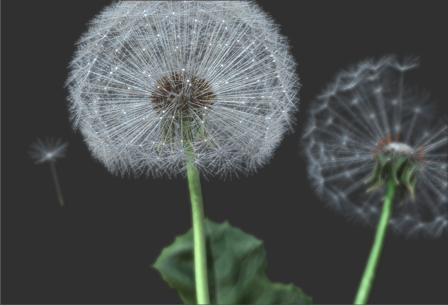
Recently I mainly work on human anatomy. I took a break and came back for a moment to botany.
Great work! And thanks for sharing your technique. 
yeah really useful
Very cool! Thanks for the breakdown. Great results!
That is easily the most bangin’ use of FiberMesh I have seen yet. Stunning! So delicate – it approaches the feeling of subtle complexity of the natural form it is modeled after.
It’s incredible!!! From viewing these works, I want to work, work, work … And learn, learn, learn …
TNX! small_orange_diamondsmall_orange_diamond
Very nice I will have to try this I’ll have to try some particles and see if I can blow them off wonder if Houdini can do something
fantastic, thanks for sharing rulonis:)
This is really cool work, thank you for sharing your workflow!
May i ask how you did the eyes of the fly? I tried to do something similar to this but i failed.
Thanks!
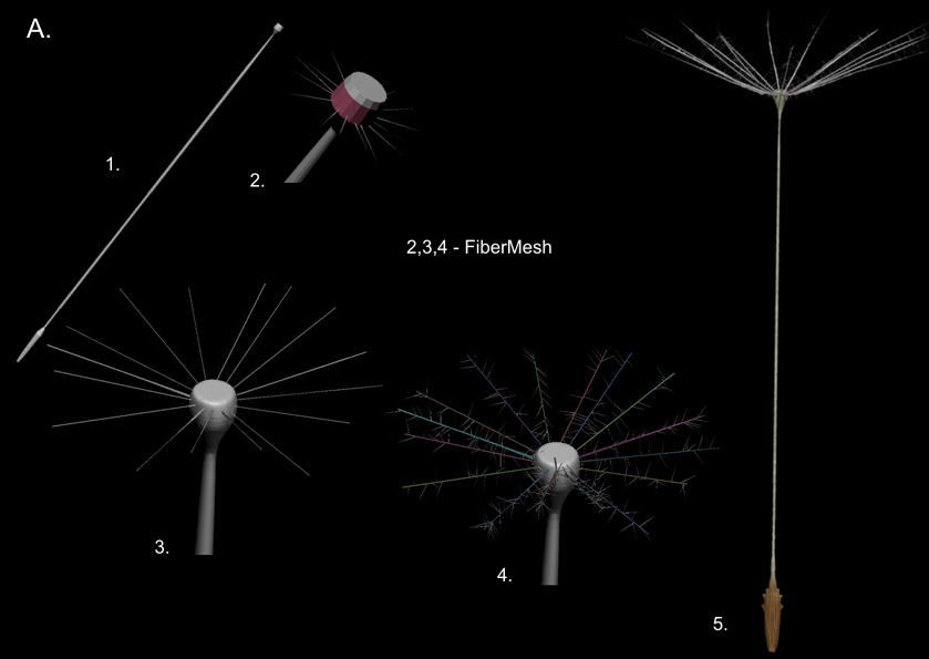
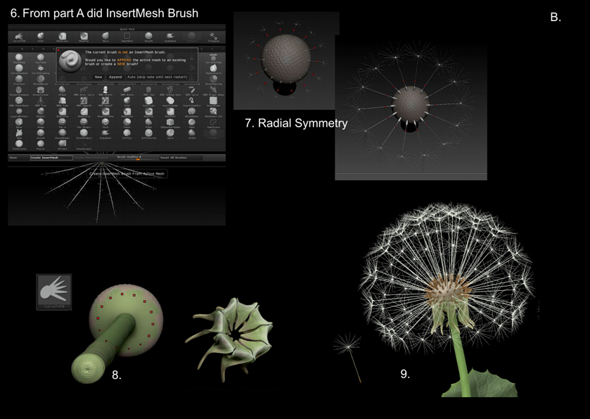
I am very glad that You liked the way how to make dandelion in ZBrush.
RahiSan I did also image of a fly, but here I think you mean bees eye .
Anyway, it does not matter here. 
Surely there are other ways, but I did it in a way that I have described in the following.
Of course, if you need to show the internal structure, then you need to make an eye with the individual elements (facets).
Super cool work, and clever use of Z
Thank you! This is very interesting!
All done in Zbrush.
Attachments
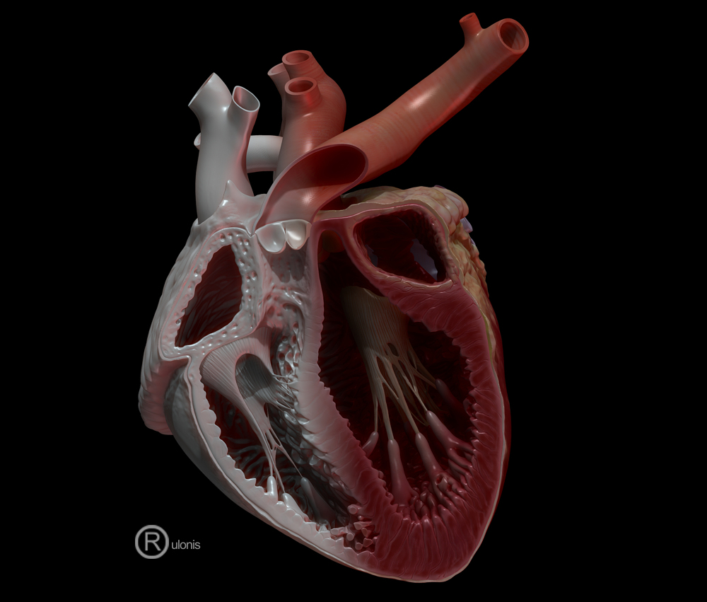
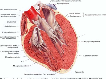
Wait there really are those thin muscle fibers in the heart ? Weird.
Anyway great models. Was teh heart all hand sculpted or did you used some 3d scan data ?
Thank you.
Yes, in the middle are the fibers that hold the valve.
Yes, it is hand sculpting.
Swietny rysownik anatom Sobotta
Heh i always imagined the heart as something solid as a rock with no fragile parts in it, guess the inside dosnt have to be so strong.
How did you get those muscle fiber flow so nicely from the side to the inside structure ? I really liek how you did it.
Awesome job on this! I’ve built a couple of harts so far, and that’s such a hard thing and really satisfying once done. I would only suggest taking a closer look of the thickness of the trabeculae carneae (walls) of the left ventricle. It is usually very thick because that’s the chamber from which the blood is pumped into the whole of the body, hence these walls (muscles) are better developed than that of the right ventricle.
Cheers!
Thank Cherub Rock. Indeed, the spatial structure of the heart is difficult for mapping. Indeed left ventricular wall may be thicker. In athletes, even very thick, but like many other characteristics a personal attribute, depending on the load of the heart. But thank you for your advice and I think I will do a little thicker.
Slocik, heart valve and the wall, of course, are two different objects. Useful was a brush SnakeHook and rebuilding the grid by DynaMesh, then mostly simple sculpting. When I did this work was not yet useful function ZRemesher
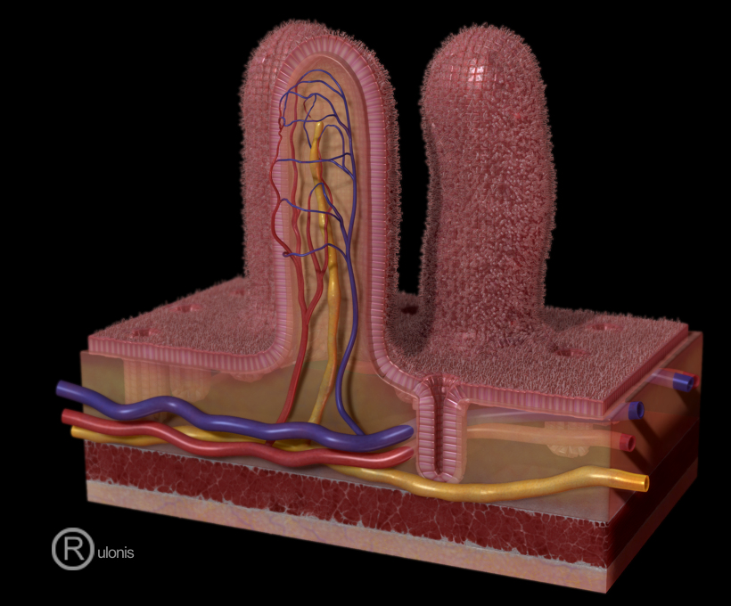
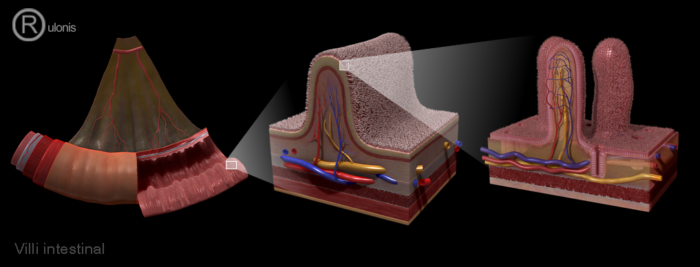
And the last little addition to showing my work on the anatomy - the small intestine. You can see the formation of cell layers and villi by using micromesh.